


| Protein: | RBP1 |
| Organism: | Homo sapiens |
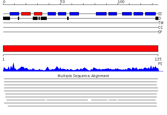
| Length: | 135 amino acids |
| Reference: | Drew K, et al. (2011) The proteome folding project: Proteome-scale prediction of structure and function. Genome Res. 2011 Sep 16 |
Listed below are up to the top 10 sequence alignment matches, by species, for the PSI-BLAST search against the protein sequence for RBP1.
| Description | E-value | Query Range |
Subject Range |
|
|
2.0E-50 | [1..135] | [63..197] |
|
|
1.0E-44 | [1..134] | [1..134] |
|
|
2.0E-44 | [1..134] | [1..134] |
|
|
2.0E-44 | [1..135] | [1..135] |
|
|
1.0E-43 | [1..135] | [1..135] |
|
|
1.0E-43 | [2..134] | [1..133] |
|
|
5.0E-43 | [1..134] | [1..134] |
|
Region A: Residues: [1-135] |
      1          11         21         31         41         51
      |          |          |          |          |          |
    1 MPVDFTGYWK MLVNENFEEY LRALDVNVAL RKIANLLKPD KEIVQDGDHM IIRTLSTFRN  60
   61 YIMDFQVGKE FEEDLTGIDD RKCMTTVSWD GDKLQCVQKG EKEGRGWTQW IEGDELHLEM 120
  121 RVEGVVCKQV FKKVQ
|
| Detection Method: | |
| Confidence: | 46.0 |
| Match: | 1jbhA |
| Description: | Cellular retinol-binding protein II (CRBP) |
Matching Structure (courtesy of the PDB): |
|
| Term | Confidence | Notes |
| long-chain fatty acid transporter activity | 5.31242454679135 | bayes_pls_golite062009 |
| fatty acid transporter activity | 5.05947161650978 | bayes_pls_golite062009 |
| lipid transporter activity | 4.30505384117861 | bayes_pls_golite062009 |
| retinoid binding | 3.86745158299736 | bayes_pls_golite062009 |
| isoprenoid binding | 3.80802424204178 | bayes_pls_golite062009 |
| lysophospholipid transporter activity | 3.53484598441971 | bayes_pls_golite062009 |
| retinol binding | 3.41359399354212 | bayes_pls_golite062009 |
| binding | 2.93830472620404 | bayes_pls_golite062009 |
| transcription regulator activity | 2.27413749831465 | bayes_pls_golite062009 |
| transporter activity | 2.15627660314839 | bayes_pls_golite062009 |
| transcription repressor activity | 1.45696778890481 | bayes_pls_golite062009 |
| protein binding | 1.23450129884619 | bayes_pls_golite062009 |
| prostaglandin-D synthase activity | 1.18764036773412 | bayes_pls_golite062009 |
| lipid binding | 1.0002255159651 | bayes_pls_golite062009 |
| retinol transporter activity | 0.811095267558536 | bayes_pls_golite062009 |
| fatty acid binding | 0.761923641769607 | bayes_pls_golite062009 |
| substrate-specific transporter activity | 0.670647472301401 | bayes_pls_golite062009 |
| retinoic acid binding | 0.539496110498594 | bayes_pls_golite062009 |
| intramolecular oxidoreductase activity | 0.50851501669551 | bayes_pls_golite062009 |
| bile acid binding | 0.37501307074598 | bayes_pls_golite062009 |