Fluorescence Microscopy
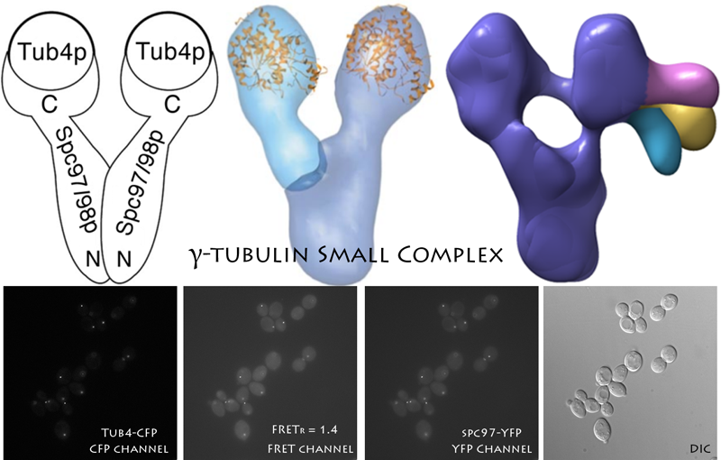
Structural analysis of the yeast gamma tubulin (TUB4) complex using FRET. See: [Kollman JM, Zelter A, Muller EG, Fox B, Rice LM, Davis TN, Agard DA. The structure of the gamma-tubulin small complex: implications of its architecture and flexibility for microtubule nucleation. Mol Biol Cell. 2008 Jan;19(1):207-15. Epub 2007 Oct 31.] and [Choy RM, Kollman JM, Zelter A, Davis TN, Agard DA. Localization and orientation of the gamma-tubulin small complex components using protein tags as labels for single particle EM. J Struct Biol. 2009 Dec;168(3):571-4. Epub 2009 Aug 31.]
The YRC develops technologies based on live-cell microscopy and FRET to map the relative position of proteins within large multi-protein complexes. The FRET information can be used for model building and is particularly powerful when paired with electron microscopy. The identification of regions in a complex that are in close proximity is also useful in the design of genetic studies. We welcome collaborations in the use of FRET to explore the structure of large protein complexes in yeast. We advise on the experimental design, and perform the microscopy and data analysis. We also train collaborators on the use of our FRET analysis software, FRETSCAL, and our FRET methodology in general. We are currently developing techniques to simplify the insertion of fluorescent proteins internally within the coding sequence of target proteins. This will make it possible to easily map the position of not only the N- and C-termini, but multiple positions along the length of a protein as well.